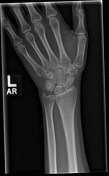
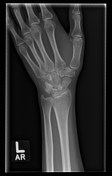
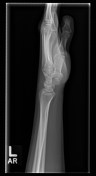
Scaphoid fracture with AVN
Diagnosis almost certain
Updates to Study Attributes
Caption
was added:
X-ray
Modality
was set to
X-ray.
Findings
was added:
Minimally displaced scaphoid proximal waist fracture with central lucency. Increased sclerosis is seen on the scaphoid view of the proximal pole of the scaphoid. No widening of the scapholunate interval. No loss of scaphoid height.
Images Changes:
Image X-ray (Frontal) ( update )
Perspective
was set to
Frontal.
Image X-ray (Oblique) ( update )
Perspective
was set to
Oblique.
Image X-ray (Lateral) ( update )
Perspective
was set to
Lateral.
Image X-ray (Scaphoid View) ( update )

Perspective
was set to
Scaphoid View.
Image 1 X-ray (Frontal) ( create )
Image 2 X-ray (Oblique) ( create )
Image 3 X-ray (Lateral) ( create )
Image 4 X-ray (Scaphoid View) ( create )
Updates to Study Attributes
Caption
was added:
CT
Updates to Case Attributes
Presentation
was changed:
Persisting left wrist and anatomical snuff box pain 8 weeks after FOOSH. A plain x-ray was unremarkable at the time of injury (not available).