Presentation
History of trauma to the left wrist 6 years ago with persistent pain.
Patient Data


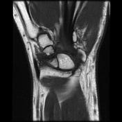
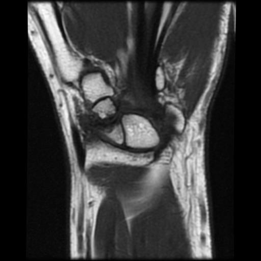
Old scaphoid fracture with non-union of the bony fragments and presence of minimal fluid within the fracture line. No evidence of osteonecrosis of the bony fragments. Narrowing of the radioscaphoid joint space with marginal osteophyte of the radial styloid and thickening with the enhancement of the radial collateral ligament.
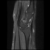
There is also mild degenerative changes of the proximal aspect of the lunate bone and in the capitate. On the sagittal sequence, the lunate bone is dorsally tilted with moderately increased lunocapitate angle at 35° (normal <30°) indicating probably a dorsal intercalated segment instability.
Case Discussion
MRI features of a scaphoid pseudarthrosis with secondary carpal instability (dorsal intercalated segment instability).


 Unable to process the form. Check for errors and try again.