

Presentation
Left neck swelling for one month. No pain, fever or discharge from the swelling.
Patient Data

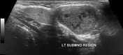
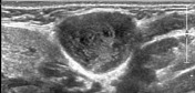
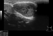
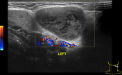
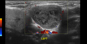
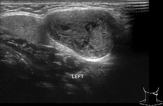
A well-defined lesion measuring about 2.6 x 1.7 cm showing mildly heterogeneous matrix and a few small cystic components, is seen along the posterior aspect of the left submandibular gland. No significant internal vascularity is seen in it.
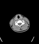

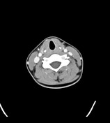
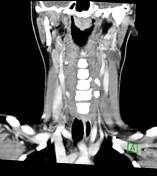
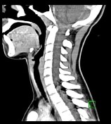
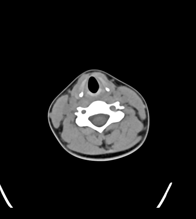
A well -defined, oval-shape cystic lesion measuring 2.6 x 2.0 x 2.0 cm is seen in the left submandibular space (posterior to the submandibular gland, anterior to the sternocleidomastoid muscle and lateral to the carotid sheath). It has mild peripheral enhancement on the post contrast study. Multiple prominent lymph nodes measuring up to 9 mm in short axis are seen at multiple levels on both sides of the neck.
Case Discussion
- well-defined cystic lesion at the left angle of mandible/submandibular space (between the submandibular gland, sternocleidomastoid muscle and carotid sheath). It is likely a 2nd branchial cleft cyst. Possible differential diagnosis includes cystic hygroma, necrotic malignant lymphadenopathy (of squamous cell carcinoma head & neck or papillary thyroid carcinoma), thymic cyst and cystic vagal schwannoma.
the lesion was surgically excised and a histopathological diagnosis of 2nd branchial cleft cyst was made.


 Unable to process the form. Check for errors and try again.