
Presentation
Abnormal antenatal ultrasound exam.
Patient Data
Age: 2 days
Gender: Female
From the case:
Semilobar holoprosencephaly


Download
Info

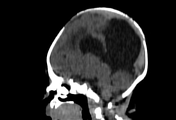
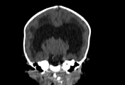
This CT scan shows:
- fused frontal lobes superiorly with grey and white matter crossing the midline
- absent anterior part of the falx cerebri
- absence of the septum pellucidum
- monoventricle with partially developed occipital and temporal horns
- prominent dorsal cyst
- incompletely formed interhemispheric fissure
- complete fusion of the thalami
- absent olfactory tracts
- hypoplasia of the corpus callosum
- hypotelorism
Additionally, the cortex appears thickened with a smooth outer surface and few poorly formed gyri. This raises the possibility of lissencephaly that could be better assessed with MRI.
Case Discussion
CT findings are consistent with semilobar holoprosencephaly with possible lissencephaly which is considered as a rare association 1. Further imaging and genetic assessment would be required to establish the lissencephaly diagnosis.
Additional contributor: A. Ramdani, MD


 Unable to process the form. Check for errors and try again.