
Presentation
Progressive shortness of breath.
Patient Data
Age: 40 years
Gender: Male
From the case:
Silicosis with progressive massive fibrosis


Download
Info
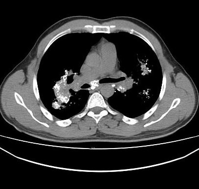
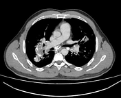
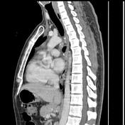
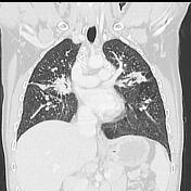
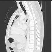
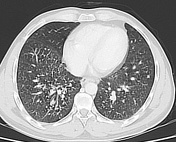
The CT scan demonstrates:
- numerous calcified hilar and mediastinal lymphadenopathy "eggshell calcification pattern"
- large mass-like conglomerates with calcifications and associated radiating strands are noted in the upper zones mainly the dorsal segment of the upper lobes, apical segment of the right lower lobe
- Multiple well-defined small lung nodules relatively uniform in shape are noted bilaterally predominantly in the upper lobes and apical segment of the lower lobes with nodular thickening of the peribronchovascular interstitium, fissural distortion and emphysematous bullae of the apical segment of the left upper lobe
- No cavitary lesion is seen
Case Discussion
The CT features of bilateral partially calcified mass-like conglomerates, mediastinal and hilar lymphadenopathy with "eggshell calcification pattern" and bilateral small lung nodules in a patient with a history of exposure to silica particles (18 years as a stone carver in this case) is highly suggestive of silicosis with progressive massive fibrosis (PMF).


 Unable to process the form. Check for errors and try again.