
Presentation
Over 1 year of left nasal obstruction and recurrent epistaxis.
Patient Data


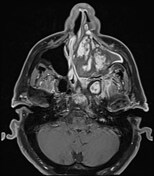
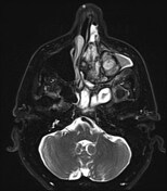
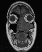
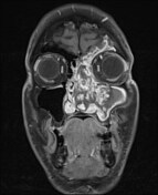
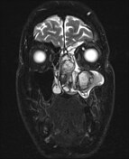
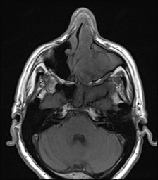
There is a 5.4 cm (AP) x 4.9 cm (TR) x 5.6 cm (CC) multilobulated, heterogeneously enhancing mass, centred at the left maxillary sinus and left nasal cavity, with rightward deviation of the nasal septum and hypointense peripheral components, which may be related to blood products. There is diffusion restriction that mostly corresponds to the enhancing components. There is associated obstruction of the left frontal and sphenoid sinuses, with T2 hyperintense mucosal thickening, and central fluid/mucus. Partial obstruction of the left ethmoid air cells.
Case Discussion
This is a case of a large sinonasal angiomatous polyp (SAP), a rare subtype of antrochoanal polyp. SAP is a proliferative fibrotic process with neovascularisation due to chronic sinus obstruction that is commonly misdiagnosed as malignancy. The diagnosis of this case was consistent with pathology by surgical biopsy:
-
left maxillary posterior wall dehiscence
focally eroded polypoid markedly inflamed granulation tissue. Negative for malignancy
-
left sinus contents
material predominantly consists of focally organising fibrin blood clots
admixed reactive mildly chronically inflamed hyperplastic and squamous metaplastic epithelium. Negative for malignancy


 Unable to process the form. Check for errors and try again.