
Presentation
Pulsatile mass in the interparietal region. Venous?
Patient Data
Age: 3 years
Gender: Male
From the case:
Sinus pericranii


Download
Info
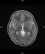
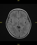
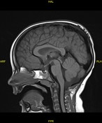
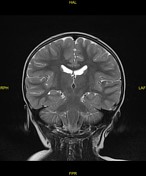
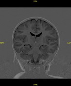
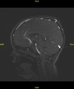
Venous communication between the intracranial superior sagittal sinuses and serpigionous epicranial veins at the vertex via a tranosseous vein.
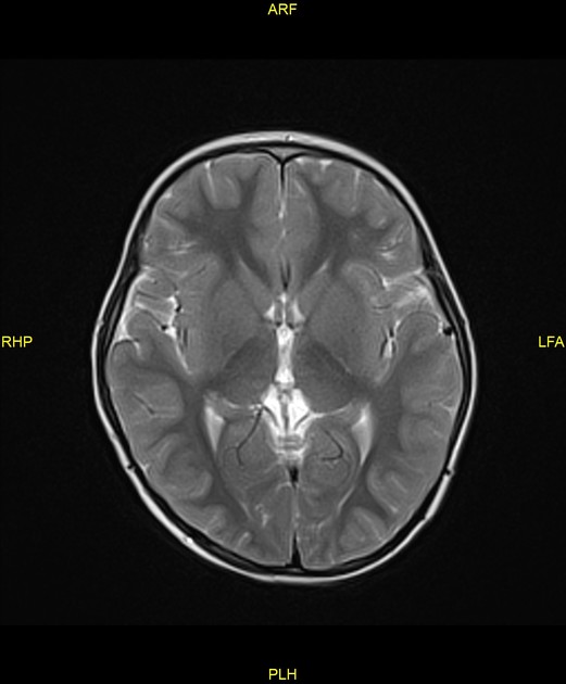
No neuroparenchymal developmental venous anomaly or AVM.
The brain is structurally normal.
Case Discussion
The MRI appearances are in keeping with sinus pericranii.


 Unable to process the form. Check for errors and try again.