
Presentation
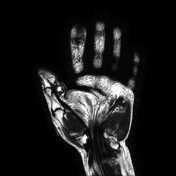
Slowly growing left thumb swelling with limitation of movement.
Patient Data
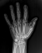

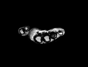
A well-defined corticated bone lesion is seen at the palmer aspect of the thumb separable from the contiguous proximal phalanx. No associated cortical destruction or bone remodeling.
Increased soft tissue thickness of the thumb and thenar regions.
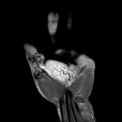

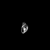
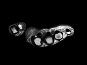

A well-circumscribed subcutaneous lesion is seen contacting the flexor tendon of the thumb. It displays high signal on both T1 and T2 images with a clearly demarcated low signal cortex. It is seen separable from the related proximal phalanx. It measures 15 x 9 x13 mm.
Subcutaneous edema of the thumb and palm is also noted.
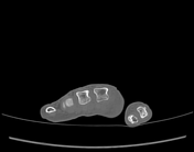
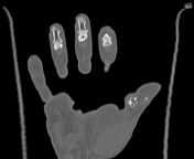


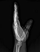
A well-circumscribed lobulated bony lesion is seen at the palmar aspect of the left thumb. It is seen associated with smooth remolding of the proximal phalanx.
Microscopic examination: revealed snips of cartilaginous matrix containing bland-looking chondrocytes with mild hypercellularity. No more than one cell/ lacuna. No myxomatous changes. No atypia, pleomorphism, or necrosis in examined material.
Diagnosis: Picture of enchondroma.
Case Discussion
Differential diagnosis includes:
osteochondroma of sesamoid bone 1
enchondroma of sesamoid bone 2


 Unable to process the form. Check for errors and try again.