
Presentation
Mobile painless mass of the upper dorsal region.
Patient Data


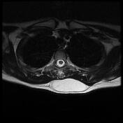
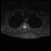
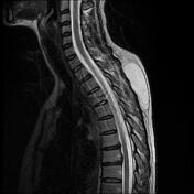
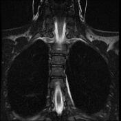
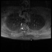
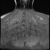
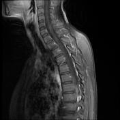
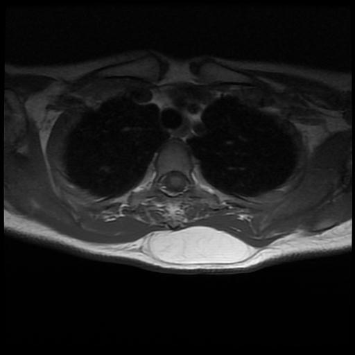
There is a well-encapsulated soft tissue mass (4 x 8 x12 cm) of the upper dorsal region from D1 to D5 levels.
It displays a high signal intensity on both T1WI/T2WI, attenuated on suppressed fat sequences (T1 fat sat and T2 fat supressed sequence), containing multiple thin fibrous septations within (< 2 mm) without obvious enhancement seen on postcontrast sequences.
Case Discussion
MRI features of an upper dorsal region soft tissue lipoma. Differential diagnoses should include normal adipose tissue ( usually non-encapsulated fatty mass, indistinguishable from the adjacent subcutaneous fatty tissue) and liposarcoma (thick enhanced septa or local invasion of the adjacent structures).


 Unable to process the form. Check for errors and try again.