
Presentation
Right proptosis.
Patient Data
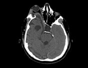

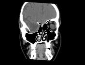
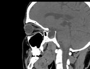
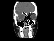
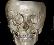
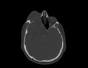
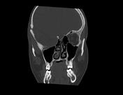

Absence of the right sphenoid wing and the lateral orbital wall. Consequently the right temporal lobe with the surrounding CSF and enlarged arachnoid space are prolapsing into the right orbit producing distorsion of the optic nerve and subsequent proptosis.
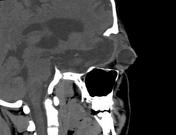
A gaping bony defect is seen in the posterior aspect of the right orbit giving the "empty orbit sign"
Absent anatomical landmarks of the optic nerve canal, anterior clinoid, superior and inferior orbital fissures.
Right upper eyelid subcutaneous soft tissue thickening, likely representing plexiform neurofibromatosis.
Diffuse atrophic changes of the right cerebral hemisphere.
Cortical irregularity of the right orbital roof and parietal bone secondary to old trauma and surgical intervention.
Case Discussion
The patient is known to have "neurofibromatosis type 1".


 Unable to process the form. Check for errors and try again.