
Presentation
Vomiting, lower limbs weakness and left eye blurred vision.
Patient Data


An ill-defined lesion is seen in the right middle cranial fossa with marked surrounding vasogenic oedema.
Mass effects are manifested as compression of underlying parenchyma, ipsilateral lateral ventricle, and mid-line shift contralaterally.
Bony changes are seen in the form of hyperostosis involving the right-sided greater wing of the sphenoid.
A note is made of empty sella.


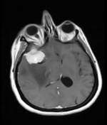


A partially circumscribed, extra-axial, altered signals intensity mass lesion is noted involving the right middle cranial fossa, the returning signals are hypo to isointense on T1, hyperintense on T2, T2 FLAIR, and show avid enhancement on post-contrast images. The lesion also shows a small internal cystic component suggesting an area of necrosis. The lesion is associated with significant perilesional oedema which extends into the right frontal region. CSF cleft and dural tail signs are seen. Mass effect is manifested by effacement of the ipsilateral lateral ventricle and underlying brain parenchyma and shifting of midline (measuring 8mm) contralaterally. The mass lesion is inseparable from the adjacent part of the greater wing of the sphenoid on the right side.
Case Discussion
CT and MRI findings along with the location of the lesion are more in favour of sphenoid wing meningioma as described above.


 Unable to process the form. Check for errors and try again.