
Presentation
Spastic lower limbs, imbalance and abnormal gait
Patient Data


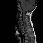
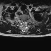

There is a large expansile and bubbly cystic bony lesion arising from the posterior element of T1 vertebra with slight extension to its body.
It appears mixed hypointense and hyperintense on both T1 and T2 with multiple fluid-fluid levels due to blood filled cavities.
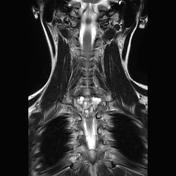
I causes marked compression of the spinal cord with severe spinal canal stenosis, but no definite compressive myelopathy. Nerve roots compression at D1-D2 level is also noted.
The rest of the spinal cord and the other visualised vertebrae appear normal. No other bony lesions.
C5-C6
Small central disc protrusion and annular tear indenting the thecal sac. No canal stenosis or nerve roots compression.
Case Discussion
Appearances in this case are highly suggestive of spinal aneurysmal bone cyst causing spinal cord compression and canal stenosis.
Aneurysmal bone cyst is a benign, vascular and locally aggressive bony lesion. It affects females more than males, and most of the cases occur in the 1st and 2nd decades.
Up to 30% of aneurysmal bone cysts are secondary to a pre-existing bony lesion.
Spine and sacrum are affected in 20 to 40 % of cases. Spinal ABCs affect mainly posterior vertebral elements with extension to vertebral bodies occur in up to 40% of cases.
Fluid-fluid levels are highly suggestive of ABC, although are not specific.


 Unable to process the form. Check for errors and try again.