Presentation
Sudden onset of upper thoracic pain and paraparesis. No trauma.
Patient Data
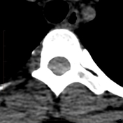
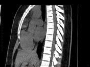
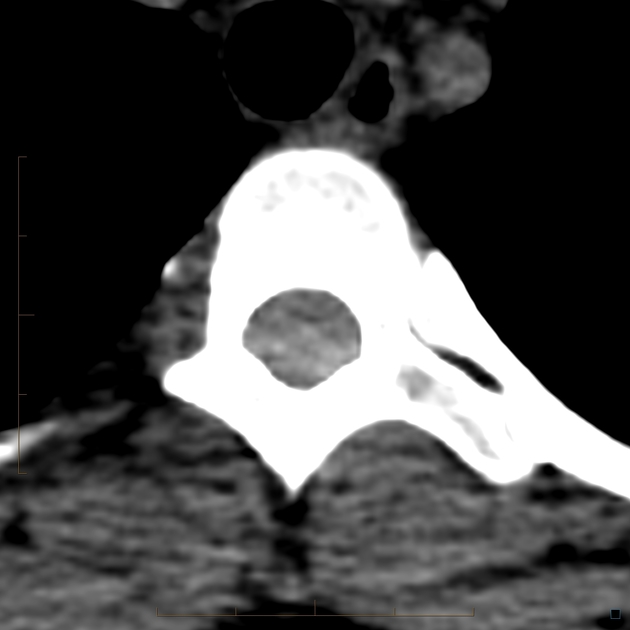
Subtle crescentic hyperdensity posterior to the spinal cord in the upper thoracic region without underlying fracture or other bone pathology.
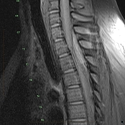

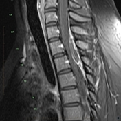

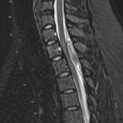

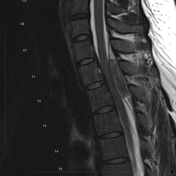


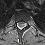

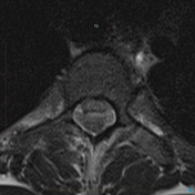

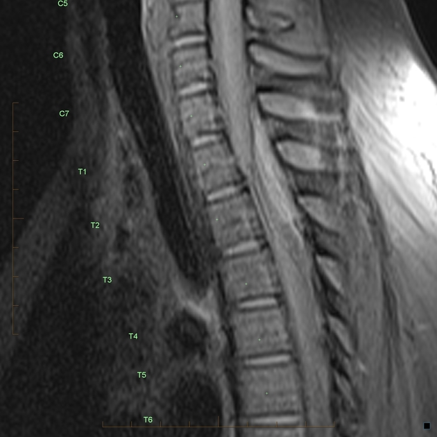
Extra-axial mass of high signal on T1 (best seen on the pre-contrast T1 fat sat axial) at the cervco-thoracic junction showing low T2 signal and peripheral enhancement. Note how the "mass" lifts up and displaces the dura on the sagittal sequences. No serpentine flow voids to indicate an underlying dural arterio-venous fistula.
Case Discussion
Spontaneous (ie non-traumatic) extradural hematoma in the spine is rare but can be devastating. They typically occur at the cervico-thoracic junction posteriorly as this is a particularly prominent site for epidural veins and often in younger persons. An underlying vascular pathology such a arterio-venous fistula may be present. The keys to diagnosis are the sudden onset of painful paraparesis/quadraparesis with an extra axial mass on MRI sitting outside the dura, of high T1 signal and low T2 signal indicative of methaemaglobin and peripheral enhacement only.