
Presentation
The patient was referred for a barium swallow study because of chest pain and odynophagia for three months.
Patient Data

A large concave impression is seen on the proximal of the thoracic oesophagus at the level of the aortic arch. In addition, a considerable mass-like opacity is evident in the upper part of the left lung in the para-mediastinal region.
A small-sized hiatal hernia is observed.
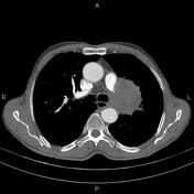


A 95 × 68 × 75 mm mass with an irregular margin, inhomogeneous enhancement and small foci of calcification is seen in the left upper lobe, which invades the adjacent mediastinum and partially encases the distal aortic arch and proximal descending part and abuts the oesophagus. Additionally, the mass extends to the left lung hilum, encases the hilar structures and obstructs the left pulmonary artery.
Mild interlobular septal thickening in the left lung, particularly around the mass, suggests lymphangitic carcinomatosis.
Case Discussion
This case demonstrates an elderly patient with chest pain and odynophagia for the past three months who was referred for a barium swallow study. Due to a large concave impression on the oesophagus at the level of the aortic arch and a sizeable mass-like opacity in the adjacent left lung, the patient underwent a contrast-enhanced chest CT on the same day.
The CT shows a huge left upper lobe lung mass that invades the mediastinum and abuts the oesophagus, corresponding to the patient's symptoms.
A bronchoscopy and tissue exam were performed for the patient's left lung mass, and histopathology evaluation confirmed squamous cell carcinoma of the lung.


 Unable to process the form. Check for errors and try again.