
Presentation
Haemoptysis and dyspnoea.
Patient Data
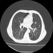

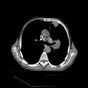

There is a 30 x 45 x 55 mm isodense solid infiltrative spiculated mass lesion involving the left upper lobe anterior and superior lingula segments with extension to the left hilum associated with adjacent tissue and parenchymal distortion making lymphangitic carcinomatosis appearance along with left upper lobe anterior segment.
Focal pleural thickening of the left upper lobe is associated with a pleural-based mass measuring about 20 x 25 x 30 mm located at the anterior aspect of LUL anterior and superior lingulae segments.
Left hilum and mediastinal lymphadenopathy, with a maximum SAD of up to 12 mm caused a pressure effect and subsequent luminal narrowing of the left upper lobe bronchus.
Diffuse emphysematous changes more dominantly with centriacinar pattern is noted.
Calcified nodule measuring about 9 mm along with left lower lobe lateral basal segment.
Two ground glass nodules with maximum diameter up to 5 mm are seen at left lower lobe.
Case Discussion
The left upper lobe mass underwent biopsy and microscopic description revealed fragments of chronically inflamed respiratory mucosa with carcinoma in situ and a small focus of squamous cell carcinoma.


 Unable to process the form. Check for errors and try again.