
Presentation
Cyst on OPG for further assessment.
Patient Data
Age: 40 years
Gender: Male
From the case:
Stafne cyst


Download
Info

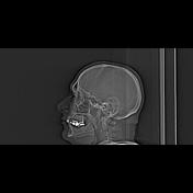
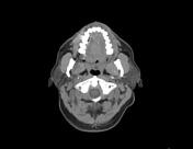
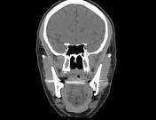
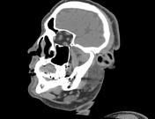
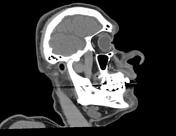
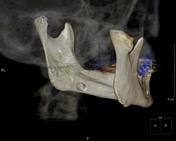
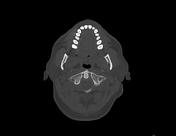
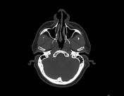
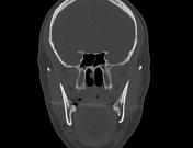
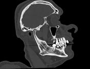
A small radiolucent well-corticated defect is seen at the lingual wall of the left mandible between the inferior alveolar nerve canal and the inferior border of the posterior mandible just anterior to the angle of the jaw. This defect is filled with fat.
Case Discussion
Stafne cysts are small cortical defects near the angle of the mandible below the mandibular canal.
Case courtesy of Dr Amir Elhamy, Theodor bilharz institute, Egypt.


 Unable to process the form. Check for errors and try again.