
Presentation
Chronic smoker, cough, breathlessness
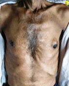
Patient Data

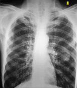
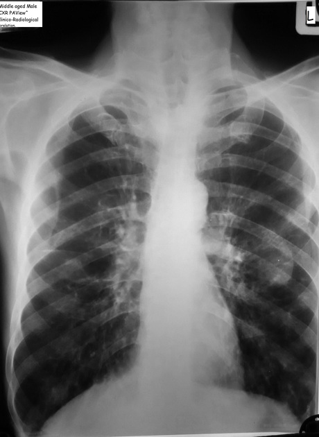
Bilateral emphysema with relatively tubular cardiac silhouette
Well defined radio-opacity in left mid zone overlapping anterior 4 and 5th ribs. no rib erosions, collapse or consolidation or abnormal calcification or air bronchogram , nor crowding of bronchovascular markings.

The well-defined lesion on the chest x-ray is not within the chest, but secondary to a subcutaneous lesion.
Case Discussion
Incidentally revealed in a middle aged patient who came for routine exam for chest evaluation because of chronic cough with breathlessness in chronic smoker (daily 12-15).
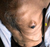
Findings on radiograph of extrinsic nature and not parenchymal lesion ; corroboration with patient history of non significant nature since long standing and clinical exam of it being soft cyst like indentable swelling since childhood with slow growth .
Take away message : sometimes clinical or radiographic pictures alone are insufficient. This information viewed together enlightens us even with limited views.


 Unable to process the form. Check for errors and try again.