
Presentation
Left leg pain and swelling after a car accident trauma a month ago.
Patient Data
Age: 80 years
Gender: Male
From the case:
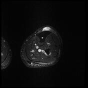
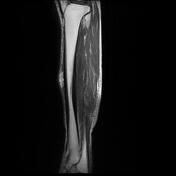
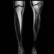
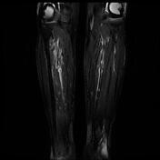
Subcutaneous hematoma - leg (MRI)


Download
Info

There is a 20 x 35 x 50 mm high signal (T1/T2) subcutaneous mass-like lesion situated anteriorly at the interface between the tibia anterior cortex and subcutaneous fat along with the middle third of the left leg.
Subcutaneous edematous changes also are seen.
Case Discussion
Regarding the imaging and clinical findings, left leg subcutaneous hematoma in the late subacute phase is the main diagnosis.


 Unable to process the form. Check for errors and try again.