
Presentation
Anterior midline neck mass slowly enlarging since childhood.
Patient Data
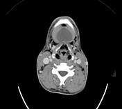

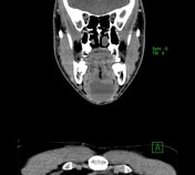
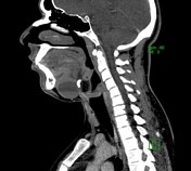
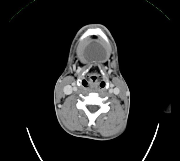
A well-circumscribed midline thin-walled cystic lesion within the submental triangle. It measures 4.2 x 3.5 x 3.5 cm (AP x TR x CC) in maximum three dimensions. It has a relatively high-density dependent contents and a posterior wall focus of calcification and no appreciable post-contrast enhancement. Appearance in keeping with complicated dermoid/epidermoid cyst.

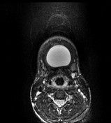
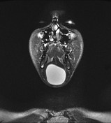
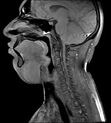

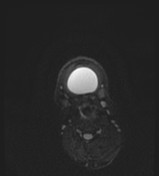
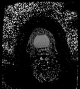
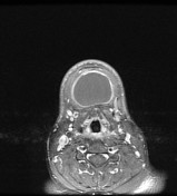
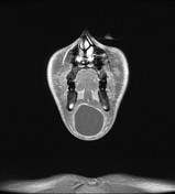
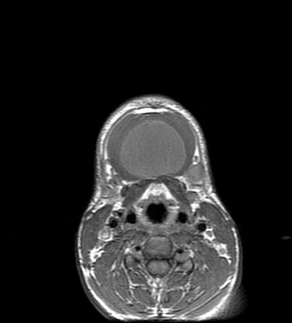
A well-defined midline thin-walled cystic lesion is seen within the submental space. It lies below the mylohyoid muscle and away from genioglossus and geniohyoid muscles, so no effect on the tongue or soft palate. It elicits diffuse homogeneous low T1W signal, high T2W signal with no drop on fat-saturation protocols or diffusion restriction as ADC shows shine-through effect. It shows uniform marginal wall enhancement in the post-contrast study. This cystic appearance has no internal septa, abnormal nodules or soft tissue.
Case Discussion
Midline cystic lesion if above the geniohyoid muscle is a sublingual swelling that will displace the tongue with consequent difficulty in eating and speaking. If the midline cystic swelling lies below the geniohyoid and mylohyoid muscles will be a submental swelling and causes double chin appearance.


 Unable to process the form. Check for errors and try again.