
Presentation
Headache, insomnia, decreased concentration, memory decline, without focal neurological symptoms.
Patient Data


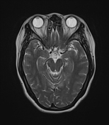
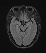
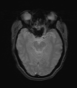
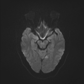
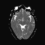
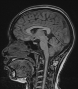
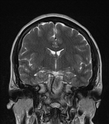
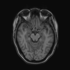
On non-contrast brain MRI, a lesion in the brain parenchyma of the left occipital lobe is present, showing high signal intensity on T2W, FLAIR, low signal on T1W, with true diffusion restriction (high on DWI and correspondingly low on ADC).
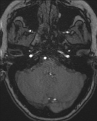
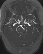
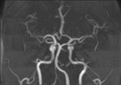
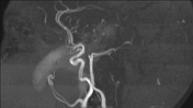
The MRA is normal without major stenosis, vascular malformation or aneurysmal dilatation.
Case Discussion
The MRI brain findings and clinical symptoms are suggestive of a subtle acute cerebral infarction. The differential diagnosis includes inflammatory and other neurodegenerative conditions.
Small, subtle lesions along with atypical clinical symptoms can easily be overlooked. DWI often assists in the early detection of these ischemic, infectious or inflammatory lesions.


 Unable to process the form. Check for errors and try again.