
Presentation
Left knee pain, trauma 8 years ago.
Patient Data

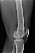
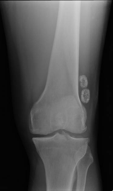
Several intra-articular calcified loose bodies
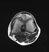

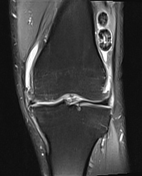
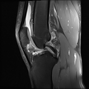
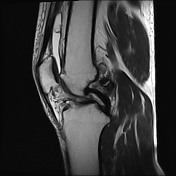

There are numerous variable-sized intra-articular loose bodies; some of them are showing intermediate to high signal intensity that is of cartilage. Others have low signal intensity at the periphery, which represents ossification.
Haziness of the ACL fibres with loss of normal signal intensity and fibre continuity suggesting a tear.
Abnormal oblique linear hyperintense signal is seen within the anterior horn of the medial meniscus disrupting its articular surface, representing a tear.
The medial collateral ligament is surrounded by oedema, with normal thickness and signal intensity of its fibres and no loss of continuit - findings are compatible with medial collateral ligament injury grade I - minor sprain.
Moderate supra-patellar effusion.
Case Discussion
The findings mentioned in this case are compatible with synovial chondromatosis. In view of the patient's age and the absence of severe degenerative process of the joint, primary synovial chondromatosis is preferred over the secondary counterpart which is a related to a degenerative process.


 Unable to process the form. Check for errors and try again.