
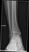
Presentation
Right medial ankle plain.
Patient Data
Age: 15 years
Gender: Male
From the case:
Talocalcaneal coalition

Show annotations
Download
Info

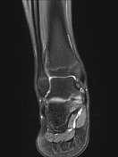
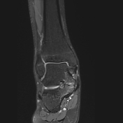
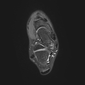
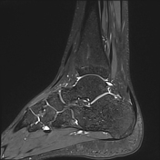
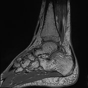
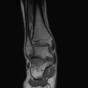
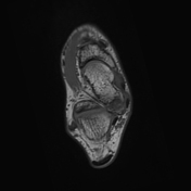
There is a fibrous/cartilaginous bridge in the medial facet of the talocalcaneal joint, with irregularity of the articular surface, narrowing of the joint space, and bone oedema, in keeping with a synchondrosis
No joint effusion. The ankle ligaments are unremarkable. Os trigonum.
From the case:
Talocalcaneal coalition
Show annotations
Download
Info
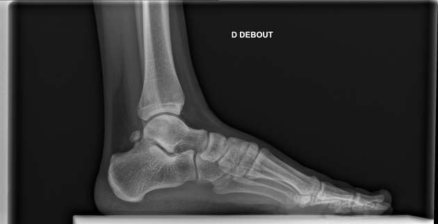
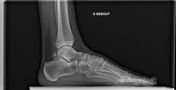
A mild continuous C-shaped arc is seen on the lateral ankle radiograph, formed by the medial outline of the talar dome and posteroinferior aspect of the sustentaculum tali due to their bridging. Os trigonum.
Case Discussion
It may be challenging to confirm non-osseous talocalcaneal coalition on plain radiographs, and an MRI is usually required to confirm the diagnosis. In fact, the C-sign has a sensitivity of almost 49%1.


 Unable to process the form. Check for errors and try again.