
Presentation
Lateral leg pain.
Patient Data
Age: 11 years
Gender: Male
From the case:
Talocalcaneal coalition - fibrous


Download
Info
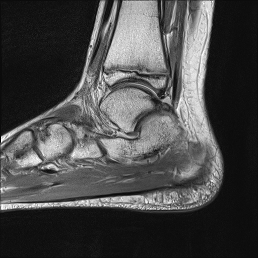
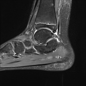
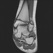
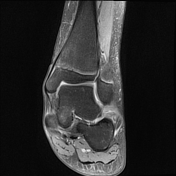
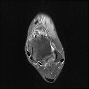
MR shows fibrous coalition between the calcaneal sustentaculum tali and the adjacent talus
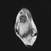
This case shows also os trigonum, peroneal and flexor muscles tenosynovitis, thickening of the superior and inferior peroneal retinacula, torn calcaneofibular ligament with attenuated anterior talofibular ligament
From the case:
Talocalcaneal coalition - fibrous
Download
Info

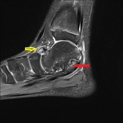
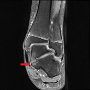
Coronal and sagittal T2WI FS fully demonstrate the TC coalition (red arrows). However, there is a talar beak, along with osseous oedema in the talus and navicular, resulting from abnormal motion at the talonavicular joint (yellow arrow), related to restricted motion at the site of talocalcaneal coalition.
Case Discussion
- tarsal coalition is 25% bilateral, even if clinical suspicion is unilateral
- calcaneonavicular (CN) ≈ 45%
- talocalcaneal (TC) ≈ 45%


 Unable to process the form. Check for errors and try again.