
Presentation
The patient presents with a complete heart block and cardiac failure. There is a new onset of dyspnea and decreased effort tolerance with ongoing syncope.
Patient Data
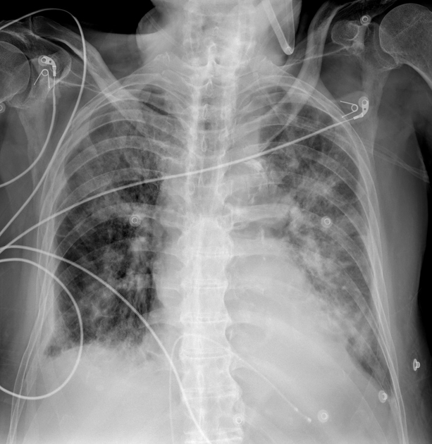
Mobile imaging performed in a cardiac ICU setting. The lung fields are reasonably inspired with ill-defined parenchymal shadowing likely representing a combination of pulmonary edema, aspiration change and possible supra-added parenchymal sepsis. There are bibasal effusions, the right greater than the left. There is an ill-defined cardiomediastinal contour, with an LV cardiac configuration. There are overlying ECG leads. There is a femoral venous access temporary single lead pacemaker terminating in the right ventricular apex.
Case Discussion
An example of a single lead, temporary cardiac pacemaker, inserted via the femoral vein with the lead terminating in the right ventricular apex. This appears low-lying however was confirmed to be intracardiac on CT (not uploaded).
In the absence of a given history, it may be difficult to realize the insertion of a temporary external pacemaker, and one can easily overlook this finding when reporting the X-ray.
The patient presents with a complete heart block, and this is a temporary emergency insertion until more definitive management is determined and afforded to the patient.


 Unable to process the form. Check for errors and try again.