
Presentation
Right testicular pain for a few weeks, getting worse over the last three days. No urinary symptoms.
Patient Data

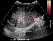
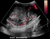
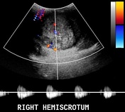
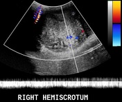
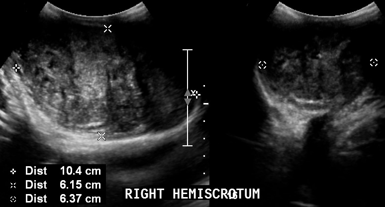
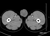
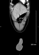
Large heterogeneous mass measuring approximately 6 x 6 x 10 cm, almost completely replacing the right testis. Color and pulsed Doppler ultrasound examinations show increased vascularity within the mass.
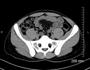

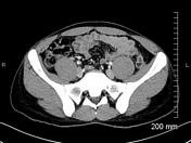
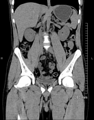
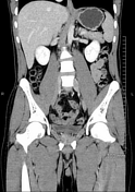
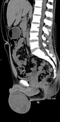
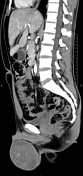
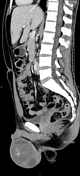
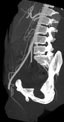
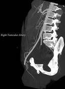
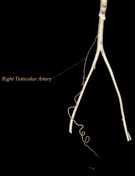

Large right testicular mass being supplied by an artery arising from the anterior aorta at L3 level between the renal and inferior mesenteric arteries (right testicular artery). It shows heterogeneous enhancement along with multiple non-enhancing cystic/necrotic areas. No abdominopelvic lymphadenopathy or distant metastasis are seen.
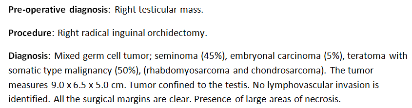
Histopathology showed mixed germ cell tumor (teratoma 50%, seminoma 45% and embryonal cell carcinoma 5%).
Case Discussion
A large heterogeneous right testicular mass was thought to likely represent a non-seminomatous germ cell tumor based on imaging findings, which was later proven to be a mixed germ cell tumor.
Laboratory investigations showed elevated tumor markers {AFP= 107 ng/ml (normal: ~8), quantitative beta-HCG =29.4 IU/L (normal in males: <5) and LDH=334 U/L (normal: 125-220)}.
The staging workup showed no evidence of distant metastasis. The patient underwent an uneventful radical right inguinal orchiectomy.


 Unable to process the form. Check for errors and try again.