
Presentation
Headache and vomiting.
Patient Data


MRI demonstrates a left thalamic lesion with no diffusion restriction and mild postcontrast heterogeneous enhancement. High choline peak is noted on MR-spectroscopy.
The lesion exerts mass effect on the third ventricle with resultant obstructive hydrocephalus.


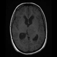
Follow up study after initiation of radio-chemotherapy demonstrates mild reduction in the size of the left thalamic lesion. The enhancing nodules are presumably from the biopsy site.
Case Discussion
Pathology:
Immunohistochemistry:
GFAP (clone polyclonal) is positive (weak)
Ki-67 (MMI) approximately 1% nuclear staining
P53 (D0-7) is positive.
Low-grade glial tumor consistent with astrocytoma WHO grade II. Sections consist with predominantly grey and white matter brain tissue with only small fragment of increased cellularity consistent with low-grade astrocytoma of WHO grade II. However, in view of the small size of the biopsy material this grade might be not representative of the actual grade of the tumor so correlation with radiological finding is mandatory.
It should be noted that the biopsy is not from the enhancing component,
located posteriorly. With such enhancement and a high Cho:Cr ratio, I
would have guessed anaplastic / Grade III astrocytoma. So there may well be, as the pathologist says, sampling error.


 Unable to process the form. Check for errors and try again.