
Presentation
The patient presented with a complaint of persistent dorsal back pain on the left side. The pain was localized mainly to the T9 dermatome and had been ongoing for several months without significant relief. She denied any history of related trauma, recent infections, or systemic symptoms such as fever or weight loss. There were no complaints of weakness, numbness, or bladder/bowel dysfunction.
Patient Data
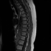

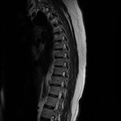
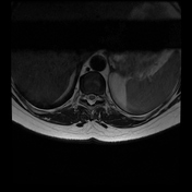
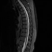
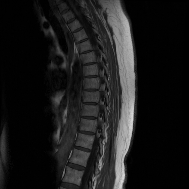
Key MRI findings:
multiple perineural cysts at various thoracic levels, largest is a cyst at the T9 foramen on the left side, measuring 1.2 cm in maximal dimension
normal bone marrow signal is seen
the vertebral bodies and disc spaces are of normal height
the cord is normal in caliber
no cavitation or abnormal signal intensities
no masses or epidural lesions
the conus ends at the regular level and is unremarkable
no disc protrusion or cord compression
no canal stenosis or foraminal narrowing
Case Discussion
Overview of perineural cysts
Perineural cysts, also known as Tarlov cysts, are cerebrospinal fluid-filled sacs. They are most commonly found in the sacral region of the spine, but they can occasionally be identified in the thoracic region. These cysts are typically asymptomatic and are often incidentally found on MRI; however, in some cases, they may be associated with radicular pain or neurological symptoms. This report discusses a case of multiple thoracic perineural cysts in a middle-aged female, emphasizing clinical presentation, imaging findings, and management.
Clinical examination
sensation: pain and temperature sensation were intact
motor function: no weakness or paresis was noted in the lower extremities
reflexes: normal deep tendon reflexes
upper motor neuron signs: Babinski and Hoffmann's signs were negative
Management and follow-up
Given the absence of neurological deficits and the lack of spinal cord compression, the patient was managed conservatively 1. She was prescribed medication for neuropathic pain relief and advised on lifestyle modifications, including postural exercises and physical therapy to reduce mechanical strain. Follow-up imaging was planned for monitoring cyst progression, and surgical intervention was not indicated at this stage 2.
Case co-authors
Dr. Zaid Khalifeh, Endo Neurosurgery Center, Amman, Jordan
Dr. Osman Elamin, Endo Neurosurgery Center, Amman, Jordan
Hind Wihaidi, School of Medicine, University of Jordan, Amman, Jordan


 Unable to process the form. Check for errors and try again.