
Presentation
History of progressive painless swelling of the upper anterior neck at midline located approximately at the hyoid bone level.
Patient Data


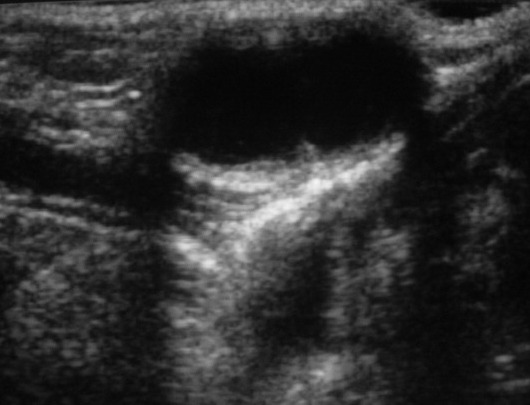
The ultrasound shows a well-defined midline cystic lesion at the level of the hyoid bone with posterior enhancement.
The transverse section at the thyroid gland shows a normal thyroid tissue in normal position.


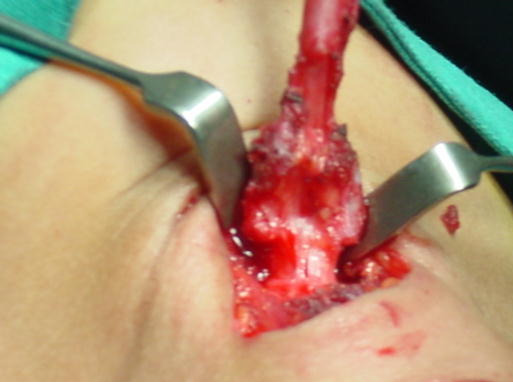
Operative view (A): excision of the cyst, duct and central part of the hyoid bone
Operative view (B): after excision
Case Discussion
Once diagnosed all thyroglossal duct cysts should be excised since infection is common making surgery more difficult, cosmetically less satisfactory and higher risk of high recurrence.
Ultrasound examination to determine the precise location of the thyroid gland is recommended since in thyroid hypoplasia, a small central area of aberrant ectopic thyroid may be mistaken for a thyroglossal duct cyst with the risk of excision and hypothyroidism.
Surgery aims to remove the entire duct including the central part of the hyoid bone to prevent recurrence.
Additional contributor: D. Ouslimane (paediatric surgeon).


 Unable to process the form. Check for errors and try again.