
Presentation
Left leg pain and limp
Patient Data
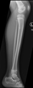
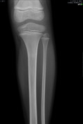
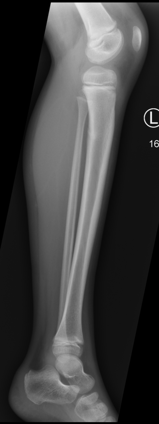
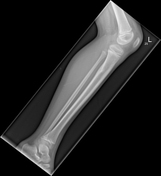
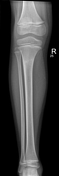
Focus of increased sclerosis at the proximal tibial metaphysis, it appears to be cortically based, arising from the posterior cortex. There is a small cortical step.
There is cortical thickening seen posteriorly in the proximal tibial diaphysis, which is associated with some defined new bone formation, that extends laterally between the tibia and fibula.


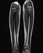
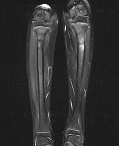
Mild osseous edema is seen on both T1 and STIR images in the proximal tibia metaphysis bilaterally. Mild thickening of the posterior cortex with periosteal reaction. Transverse low signal trabecular fracture is seen bilaterally, but now more pronounced on the right-side.


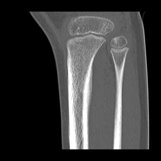
There is symmetrical cortical thickening at the posterior aspect of the proximal tibial metaphysis bilaterally.
Case Discussion
Bilateral tibial insufficiency fractures are demonstrated with the fracture in the right leg picked up incidentally at the time of MRI which was performed due to concern of possible osteoid osteoma.
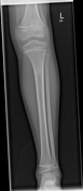
Follow-up X-ray 3 months later demonstrated stable appearances and the patient recovered fully with conservative measures.


 Unable to process the form. Check for errors and try again.