
Presentation
Primary hyperthyroidism
Patient Data
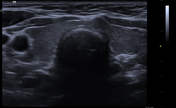
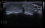
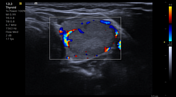
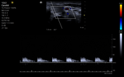

A well-defined isoechoic solid nodule in the left lobe of the thyroid at the upper pole with marked intralesional vascularity.
The rest of the thyroid gland appears normal.
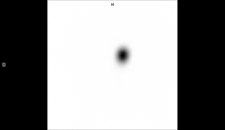
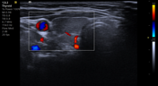
Markedly increased perfusion and tracer uptake are noted in the upper pole of the left lobe of the thyroid gland, with no significant tracer uptake in the right lobe and the rest of the left lobe—suggestive of toxic adenoma.
Case Discussion
Toxic adenoma is one of the common causes of primary hyperthyroidism. The ultrasound findings of toxic adenoma are non-specific. Thyroid scintigraphy is the modality to diagnose toxic adenoma.
Case co-authors:
Dr Mrityunjay Kumar Singh (MD, Internal Medicine), Medanta Hospital, Patna, India.
Dr Kumar Gaurav (DNB, Nuclear Medicine), Medanta Hospital, Patna, India.


 Unable to process the form. Check for errors and try again.