
Presentation
Lower anterior neck swelling.
Patient Data
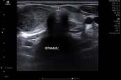
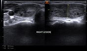
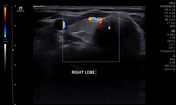
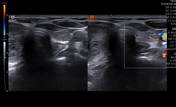
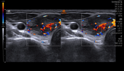
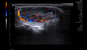
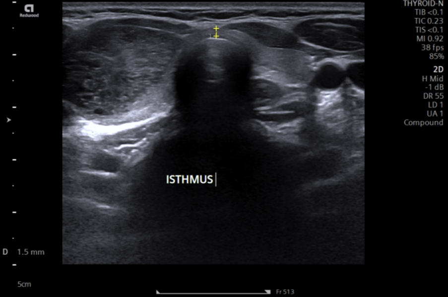
The well-defined heterogeneously isoechoic solid lesion in the right lobe of the thyroid has marked internal vascularity (TR3).
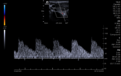
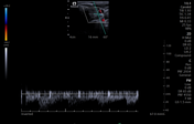
The right inferior thyroid artery shows increased peak systolic velocity (~70-80 cm/s).
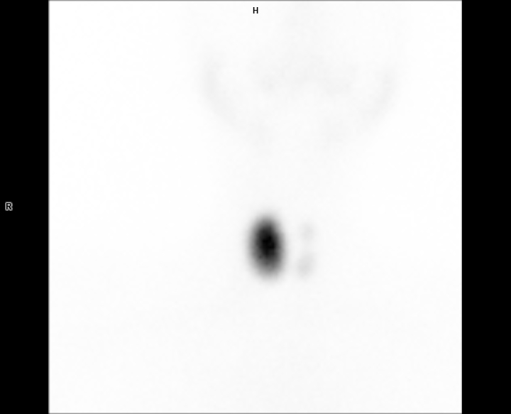
The thyroid scan shows increased uptake corresponding to a right lobe thyroid lesion.
Case Discussion
During the patient's routine investigation, primary hyperthyroidism was identified. In the context of primary hyperthyroidism, the right thyroid lesion with marked internal vascularity and increased peak systolic velocity in the ipsilateral inferior thyroid artery is most likely a toxic adenoma. The thyroid scan confirmed the ultrasound findings.
Case co-author: Dr. Mrityunjay Kumar Singh, MD (Internal Medicine), Medanta Hospital, Patna, India.


 Unable to process the form. Check for errors and try again.