
Presentation
Shortness of breath.
Patient Data
Age: 80 years
Gender: Male


Download
Info

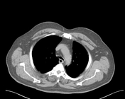
Axial CT scan (lung and mediastinal windows) shows a collapsed trachea with a decrease in the AP diameter involving the intra-thoracic part of the trachea. Segmental left lower lobe passive collapse that is related to the pericardial fat pad was noticed.
Case Discussion
This patient sent for CT scan as a patch of consolidation seen in the left lower zone of the frontal CXR that seem to be related to the prominent fat pad. The scan, for a moment of good luck, taken in the expiration so the finding of tracheomalacia became more obvious. Tracheomalacia is still either incidental or the cause of the dyspnea.


 Unable to process the form. Check for errors and try again.