
Presentation
Right flank pain with haematuria. No fever, vomiting or weight loss.
Patient Data
Atrophic right kidney with increased parenchymal echogenicity and moderate hydronephrosis. Normal looking left kidney with mild compensatory hypertrophy.
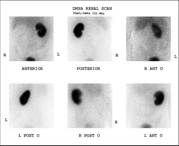
No radiotracer activity is noted in the region of right kidney (non-functioning right kidney). Left kidney shows homogeneous radiotracer uptake.


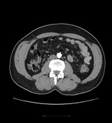
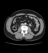
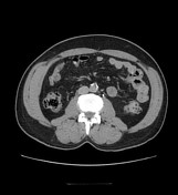
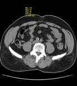
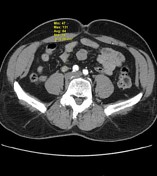
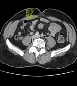

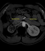
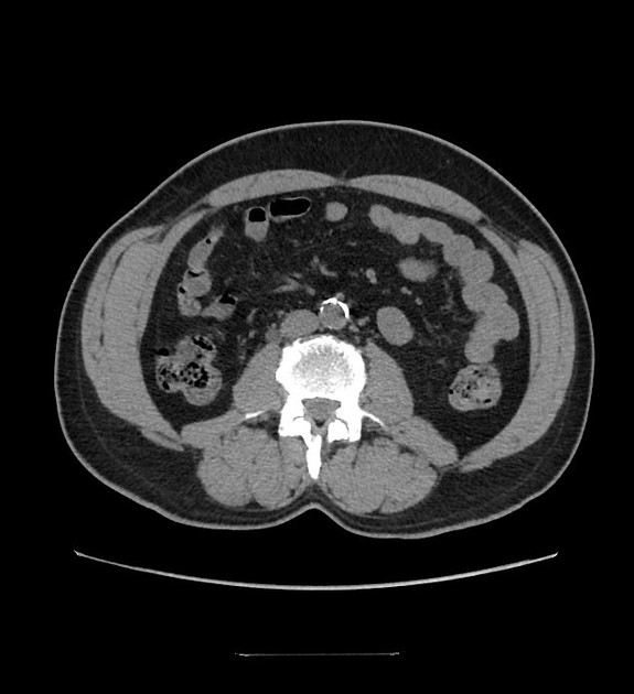
Findings: Relatively small right kidney with decreased cortical thickness, moderate hydronephrosis & mild perinephric fat stranding. Relatively small right renal artery. Dilated tortuous right ureter up to the right sacroiliac joint where there is a zone of transition and distal to this the ureter is of normal calibre. A focal abnormality measuring approximately 5.5 cm in craniocaudal length, having an average density of 30, 84, 86 & 81 HUs on plain, arterial, venous & renal excretory phases respectively, is seen in the right ureter (L3--L5 level). Left kidney, ureter and urinary bladder are grossly normal. Mildly bulky lateral limb of left adrenal gland. Impression: Right ureteric enhancing lesion suspicious of transitional cell carcinoma (TCC) with proximal hydroureteronephrosis and relatively small right kidney.
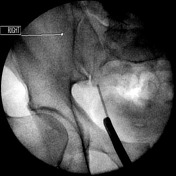

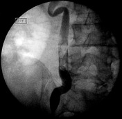
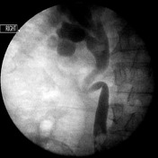
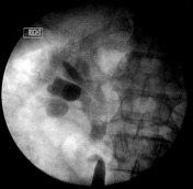
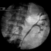

Cystoureteroscopy with retrograde pyelography
Moderate right hydronephrosis and hydroureter with a zone of transition at the level of sacroiliac joint. A filling defect (likely a mass lesion) is seen in the right ureter (L3-L5 level). Normal looking left renal pelvicalyceal system & ureter.
Case Discussion
Urine analysis: Atypical transitional epithelial cells, highly suspicious of carcinoma.
Ureteroscopic biopsy: Inconclusive due to crushing artifacts.
Procedure: Laparoscopic right nephrouretrectomy.
Diagnosis: Low grade papillary transitional cell carcinoma of the right ureter. Tumour maximum size 3.0 cm (gross). All ureteric excision margins free of tumour. Tumour does not invade submucosa or muscle coats. Negative for lymphovascular space invasion. Kidney shows severe interstitial chronic nephritis. Hydroureteronephrosis grossly confirmed. Renal vessels show severe intimal fibrosis and luminal narrowing. Renal vascular resection margin free of tumour. Pathological staging pTa,pNx, pMx.


 Unable to process the form. Check for errors and try again.