
Presentation
Presented to ED following a fall.
Patient Data
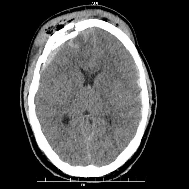
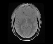
Brain:
Right frontal extradural haematoma with overlying frontal bone fracture.
Left frontoparietal and right parafalcine subdural haematoma.
Round intra-axial haematoma in the right frontal white matter, and another similar hyperdense haematoma in the left posterior temporal lobe, cortically based. Several other smaller hyperdensities scattered throughout the left cerebrum at the subcortical region and right internal capsule anterior limb/genu. These are suspicious for haemorrhagic DAI.
Crowded basal cisterns suggestive of early transtentorial herniation.
Fractures:
Open comminuted and elevated right frontal bone fracture, decompressing the increased intracranial pressure. Superiorly, the diploic spaces are separated. Fracture extends to the right frontal sinus, orbital roof and squamous part of the temporal bone.
Skull base: Non displaced left clivus fracture. Comminuted sphenoid sinus fracture. Bilateral non displaced occipital base fractures. Right sided fracture extends to the jugular foramen and towards, but not involving, occipital condyle
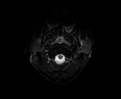
Right orbit: Minimally displaced medial and lateral walls wall. Orbital roof fracture with a gap. Nondisplaced orbital floor/maxillary sinus roof fracture. Extraconal haematoma with resultant proptosis.
Right maxillary sinus: Separated anterior wall fracture, which tranverses vertically inferiorly to involve the alveolar process. Fracture lies between 12 and 13 teeth. Depressed posterior wall fracture. Nondisplaced roof fracture extending to the lateral wall.
Bilateral comminuted and depressed nasal bone fracture. Nasal bone impacted into the ethmoid bone. Nondisplaced right zygomatic arch fracture.


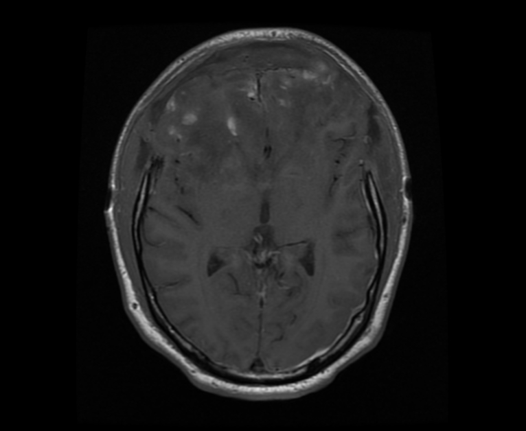
Bilateral frontal craniectomy with outward herniation of frontal lobe parenchyma related to extensive intracranial contusions and oedema.
There is involvement of the right caudate head and anterior limb of the right internal capsule as well as the genu of the corpus callosum. Multiple areas of less prominent contusion and oedema within the left posterior temporal lobe, bilateral mesial temporal lobes anteriorly and the right cerebellar hemisphere.
Involvement of corpus callosum body and dorsal lateral midbrain on the right suggests a component of sheering mechanism (diffuse axonal injury) in addition to direct compressive contusion.
Case Discussion
Rehabilitation notes
As observed in the scans the main lesion involved bilateral frontal lobes. Lesion territory included the prefrontal cortex rather than the precentral cortex thus affecting executive functions with relatively preserved motor functions.
Of additional importance is the right basal ganglia lesion due to its common association with the development of muscle spasticity1. Spasticity can appear early2, contribute to the development of contractures and adversely affect motor recovery.
After 8 months of altered consciousness rehabilitation commenced. The patient achieved independent mobility despite the early development of extensive spasticity. Executive functions were his main deficits especially poverty of thought, reduced motivation, initiation and volition. Therefore despite achieving good motor recovery he remained cognitively dependent.


 Unable to process the form. Check for errors and try again.