Traumatic urethral transection
Updates to Study Attributes
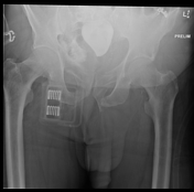
The foleyFoley catheter with a balloon is inflated and lays in the fossa navicularis, around the level of the bulbous urethra diaphragm. There is an abrupt cut-off. There is with extraperitoneal contrast extravasation and. There is no evidence of it extendingextension into the posterior urethra or the bladder.
This is evident of These findings represent a traumatic urethral transection.
Residual contrast in the right hemi pelvis on the first image is due to a previous unsucessful contrast study attempt.
Image X-ray (Preliminary ) ( update )
Updates to Study Attributes
Multiple left-sided rib fractures, left clavicle fracture, left 9th transverse process fracture, and a small left haemopneumothorax with left ICCintercostal drain in situ.
High-grade stenosis of the coeliac trunk with minor poststenotic dilatation
Complex pubic rami and left sacral ala fracture with evidence of blood within the urinary bladder and extensive pelvic haematoma. Prominent enhancement at the left base of the penis may represent a vascular injury, however there is no significant change on portal venous and delayed phases. This injury has high correlation with urethral injury.
Updates to Case Attributes
Traumatic urethral transection secondary to pelvic fractures are not common but, however havewhen present, are associated with significant implications for the patientmorbidity.
The injury occurs most commonly at the level of the bulbomembranous junction, they are not caused by sharp edges of fracture fragments rather as a result of ligamentus rupture during pelvic-ring trauma.
-<p>Traumatic urethral transection secondary to pelvic fractures are not common, however have significant implications for the patient. The injury occurs most commonly at the level of the bulbomembranous junction, they are not caused by sharp edges of fracture fragments rather as a result of ligamentus rupture during pelvic-ring trauma.</p>- +<p>Traumatic urethral transection secondary to pelvic fractures are not common but, when present, are associated with significant morbidity.</p><p>The injury occurs most commonly at the level of the bulbomembranous junction.</p>
References changed:
- Alwaal A, Zaid UB, Blaschko SD, Harris CR, Gaither TW, McAninch JW, Breyer BN. The incidence, causes, mechanism, risk factors, classification, and diagnosis of pelvic fracture urethral injury. (2015) Arab journal of urology. 13 (1): 2-6. <a href="https://doi.org/10.1016/j.aju.2014.08.006">doi:10.1016/j.aju.2014.08.006</a> - <a href="https://www.ncbi.nlm.nih.gov/pubmed/26019970">Pubmed</a> <span class="ref_v4"></span>
- 1. Alwaal A, Zaid UB, Blaschko SD et-al. The incidence, causes, mechanism, risk factors, classification, and diagnosis of pelvic fracture urethral injury. Arab J Urol. 2015;13 (1): 2-6. doi:10.1016/j.aju.2014.08.006 - Free text at pubmed - Pubmed citation