
Presentation
Right hemifacial numbness.
Patient Data
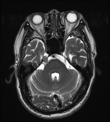

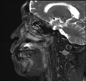
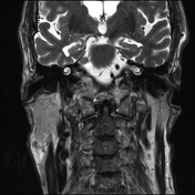
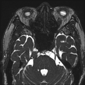
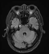
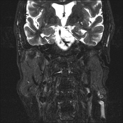
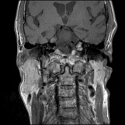
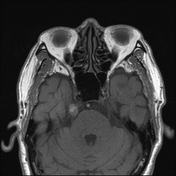
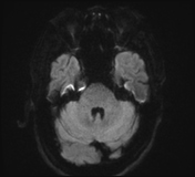
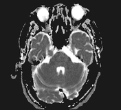
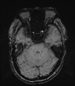
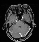
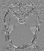
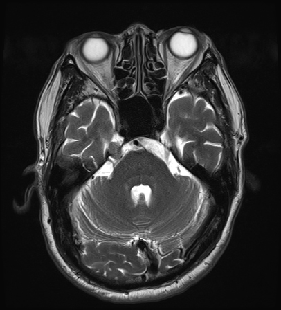
A nodular lesion involving the right trigeminal nerve (cranial nerve V) at the cisternal segment and Meckel's cave segment, measuring approximately 9 × 13 × 7 mm, showing high signal intensity on T1W and FLAIR, intermediate signal on T2W, no diffusion restriction, and strong homogeneous enhancement after contrast administration, with no significant mass effect observed.
Scattered nodular and patchy lesions in the subcortical white matter, deep white matter of the centrum semiovale, periventricular regions, and bilateral corona radiata, suggestive of cerebral small vessel disease.
Case Discussion
Imaging findings suggest a Meckel cave tumour. Due to its small size and localised nature, the most common diagnosis remains trigeminal schwannoma. The patient had no prior history of primary malignancy. Differential diagnoses include meningioma and neurolymphomatosis.


 Unable to process the form. Check for errors and try again.