
Presentation
Left ankle twist. Unable to bear weight with a tender ankle.
Patient Data

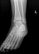
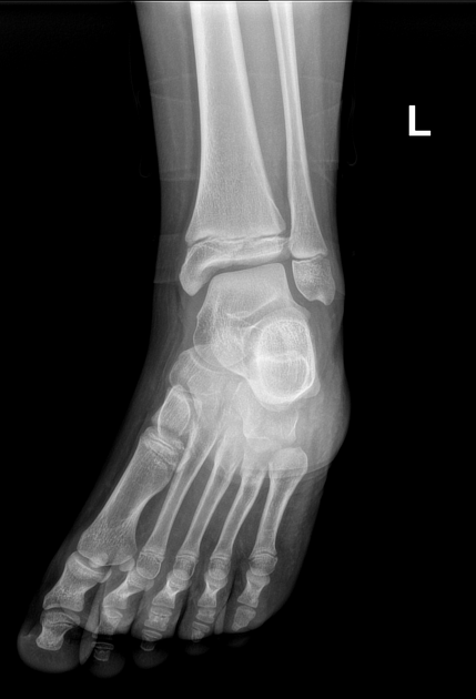
On AP view, there is vertical fracture through the lower tibial epiphysis extending to physeal plate (Salter-Harris type III appearance).
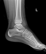
On lateral view, there is a vertical (coronally orientated) fracture through the lower tibial metaphysics extending to physeal plate (Salter-Harris type II appearance).


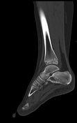
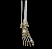
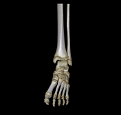
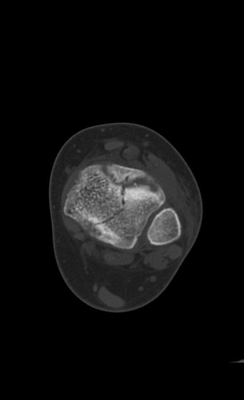
Distal tibial fracture involving the epiphysis (sagittal plane), physis (transverse plane) and metaphysis (coronal plane). It reaches the articular surface with no displacement.
Mild ankle joint effusion with peri-articular oedema.
Case Discussion
Plain radiograph and CT appearance of a distal tibial physeal injury extending to epiphysis and metaphysics in three different orthogonal planes, known as Triplane fracture (Salter-Harris type IV fracture). It usually occurs in older children aged 12 to 15 years, as the medial portion of the growth plate is closed, while the lateral portion is still open.


 Unable to process the form. Check for errors and try again.