
Presentation
History of facial trauma with swelling and tenderness over left zygoma.
Patient Data


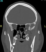
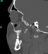
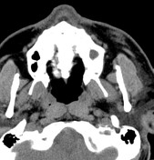
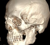
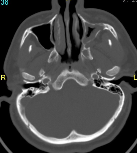
Multiple fracture lines are seen involving the anterior and lateral wall of the left maxillary sinus extending to the inferior orbital rim, additionally diastasis of the left zygomatico-frontal suture extending to the lateral orbital rim and fractured left zygomatic arch. The aforementioned fractures lead to a rather complete separation of the left zygoma with no significant displacement or facial asymmetry.
Fractured left maxillary lateral boundaries are seen radiating to the left pterygoid plate.
Partial opacification of the left maxillary sinus with mucosal thickening.
Case Discussion
Tripod fracture or zygomaticomaxillary complex fractures are comprised of fractures of the:
- zygomatic arch
- inferior orbital rim, and anterior and posterior maxillary sinus walls
- lateral orbital rim


 Unable to process the form. Check for errors and try again.