
Presentation
Seizures.
Patient Data


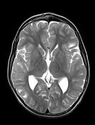
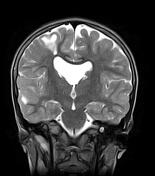
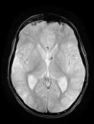
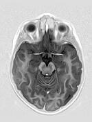

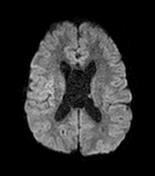
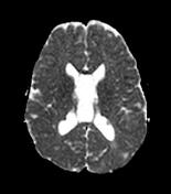
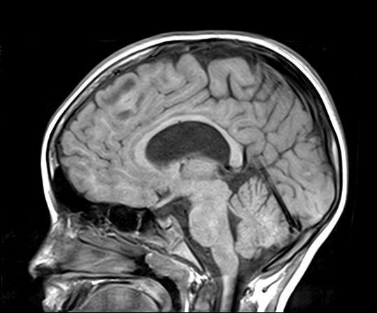
The MRI sequences demonstrate:
multiple bilateral cortical and subcortical areas of low signal on T1, high signal on T2 and FLAIR with no enhancement on postcontrast sequences in keeping with cortical/subcortical tubers.
cluster of tiny cysts of CSF signal in the white matter arround the trigone and anterior horn of the lateral ventricles with surrounding abnormal high signal on FLAIR and T2, representing probably a cystic degeneration
numerous small subependymal nodules (hamartomas) along the wall of the lateral ventricles, iso-to high signal to the grey matter on T1, isosignal on T2 and FLAIR with moderate enhancement on postcontrast sequences. Some of them show a low signal on GE (calcified nodules)
Cavum septum pellucidum and vergae are noted (incidental findings)
Case Discussion
MRI feartures of tuberous sclerosis (known case on follow-up).


 Unable to process the form. Check for errors and try again.