
Presentation
Seizures with intellectual disability and facial adenoma sebaceum.
Patient Data


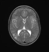
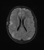
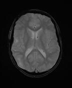
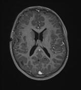
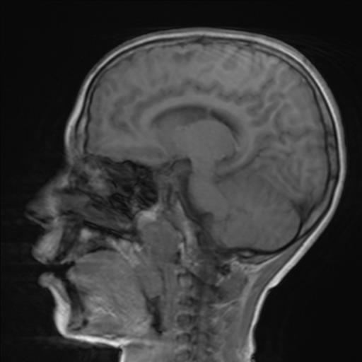
The MRI sequences demonstrate:
multiple bilateral cortical and subcortical areas iso-to-hypointense to the cortical grey matter on T1, hyperintense on T2 and FLAIR fat sat with no enhancement on postcontrast sequences in keeping with cortical/subcortical tubers.
numerous small subependymal nodules (hamartomas) along the wall of the lateral ventricles, iso-to hyperintense to the grey matter on T1, isosignal on T2 and FLAIR with moderate enhancement on postcontrast sequences. Some of them show a low signal on GE (calcified nodules)
a linear band of high signal on T2 and FLAIR fat sat is noted in the right frontal region, extending from the periventricular white matter to the subcortical region corresponding to the "radial bands sign", also known as "radial migration bands"
Case Discussion
The clinical presentation with the pathognomonic triad (Vogt triad) and the MRI exam with two major features (subependymal nodules and radial migration line) make a definite diagnosis of tuberous sclerosis.


 Unable to process the form. Check for errors and try again.