
Presentation
Seizures.
Patient Data
Age: 20 years
Gender: Male
From the case:
Tuberous sclerosis


Show annotations
Download
Info

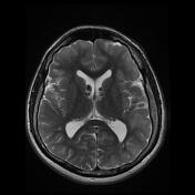
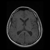
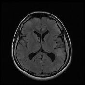
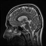
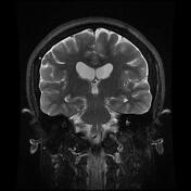
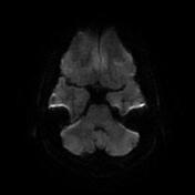
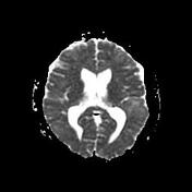
extensive T2/FLAIR cortical and subcortical white matter regions of hyperintensity due to cortical and subcortical tubers
subependymal nodules (hamartomas), which demonstrate a low T2/FLAIR signal in keeping with calcification
multiple hyperintense bands radiating from the periventricular region to the cortical surface (radial band sign)
Case Discussion
This case demonstrates typical MRI findings of tuberous sclerosis of the brain with cortical and subcortical tubers, calcified sub-ependymal nodules, and hyperintense radial bands.


 Unable to process the form. Check for errors and try again.