Tuberous sclerosis
Diagnosis possible
Updates to Study Attributes
Findings
was added:
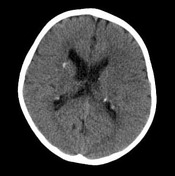
Non-contrast CT demonstrates multiple calcified subependymal nodules along the margins of the lateral ventricles. Numerous low-density regions are also seen through the subcortical white matter in keeping with cortical tubers.
Images Changes:
Image CT (non-contrast) ( update )

Perspective
was set to
Axial.
Specifics
was set to
non-contrast.
Image CT ( update )
Description
was removed:
Single Or Stack Root
was set to
.