
Presentation
Pelvic pain.
Patient Data


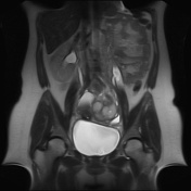
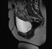
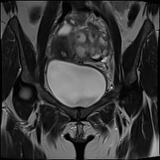
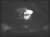
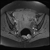
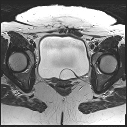
A left intra-ovarian cystic lesion with low signal intensity on T1 and intermediate signal on T2 with diffusion restriction, suggestive of left ovarian abscess.
There is a tubular structure posterior and medial to the left ovarian cystic lesion with the same MRI signal features, suggestive of left pyosalpinx.
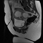
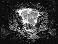
Duplex left renal collecting system with thinned parenchyma of the upper moiety and mild hydroureteronephrosis, more affecting the upper moiety.
The upper moiety ureter ends with a ureterocele which opens inferomedial to the lower moiety orifice.
Case Discussion
Features are impressive of left tubo-ovarian abscess and left pyosalpinx with an incidental note of left renal duplex collecting system with a ureterocele.
According to Weigert-Meyer law in duplex kidney with complete ureteral duplication, the upper renal moiety ureter has an ectopic insertion inferomedial to the lower renal moiety ureter and frequently ends in a ureterocele and is more prone to obstruction with resultant hydroureteronephrosis that can lead to partial atrophy of the upper renal moiety (as in this case).


 Unable to process the form. Check for errors and try again.