
Presentation
Seizures.
Patient Data


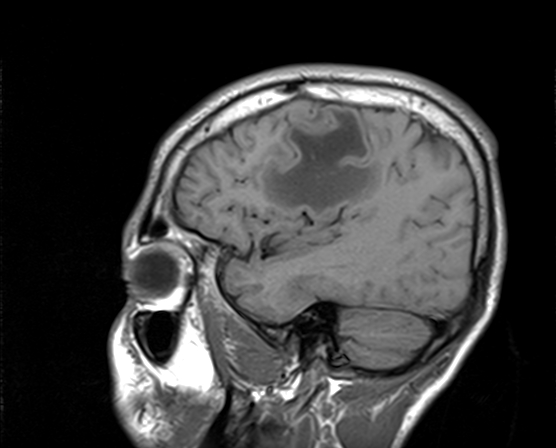
There is a large well-defined lobulated right frontal lesion, located in the subcortical white matter (subcortical U-fibers) and centrum semiovale of low signal intensity on T1WI, and high signal intensity on T2WI/FLAIR. The postcontrast sequence shows a characteristic enhancement in the form on an open-ring, the incomplete portion of the ring is on the grey matter side of the lesion. No significant perilesional edema or mass effect seen. The diffusion imaging shows an increased ADC centrally with restricted diffusion peripherally which can help to differentiate it from brain abscess in which there is a restricted diffusion.
The MRS (TR=1323, TE=136) shows elevated choline with prominent lactate (doublet).
No other white matter lesion seen.
Case Discussion
MRI features suggestive of a large tumefactive demyelinating lesion.
Additional contributor: C. Boukaaba, MD


 Unable to process the form. Check for errors and try again.