
Presentation
Patient presented to the emergency room with a one week history of left lower limb weakness with gait difficulty and recurrent tripping. Patient also notes a few day history of left hand weakness.
Patient Data


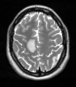
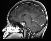
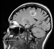

There is a 1.5 x 1.2 x 1.1 cm ring enhancing mass in the white matter of the right posterior frontal lobe. There is ring enhancement and mild oedema with no significant mass effect on surrounding structures.
Case Discussion
The MRI brain findings, along with the clinical presentation, raised suspicion for a primary neoplasm. She had no prior history of malignancy and a CT thorax abdomen pelvis revealed no lesions. A brain biopsy and lumbar puncture were performed (results below).
Pathology results
brain biopsy: hypercellular brain parenchyma with abundant macrophages, dense perivascular inflammatory infiltrates, and reactive astrocytes
lumbar puncture: myelin basic protein elevated, zero oligoclonal bands
Differential
CNS lymphoma
tumefactive multiple sclerosis
stroke
Plan
repeat MRIs to evaluate for dissemination in time and dissemination in space
serial lumbar punctures to rule out CNS lymphoma
Of note, she received IV dexamethasone for 1.5 weeks before the brain biopsy, which could have affected the biopsy results.
Key learning points
although pathology supports a demyelinating process, the absence of oligoclonal bands and other white matter lesions does not match this conclusion
the brain biopsy showed no signs of CNS lymphoma but this must be reviewed knowing that the patient had already received 1.5 weeks of IV dexamethasone


 Unable to process the form. Check for errors and try again.