
Presentation
Swelling right upper medial thigh for 3 months. Gradually increasing in size, and associated with intermittent pain. No history of trauma, fever, or weight loss.
Patient Data
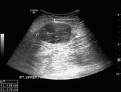
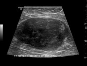
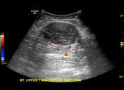
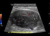

a well-defined oval shape mildly heterogeneous hypoechoic mass lesion is seen between the muscle layers of the right upper medial thigh. Mild internal vascularity is seen in it on colour Doppler ultrasound examination
differential diagnosis includes peripheral nerve sheath tumour (like neurofibroma), or soft tissue tumour (myxoma, or soft tissue sarcoma)
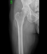
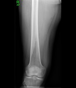
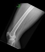
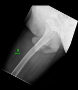
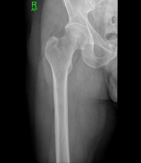
A partially circumscribed soft tissue swelling/bulge is seen along the medial aspect of the right upper thigh. No soft tissue calcifications are seen. Morphology of the visualised bones is intact.
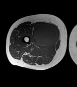

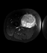
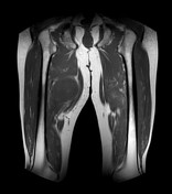
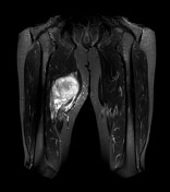
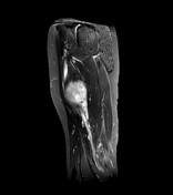
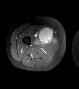
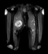
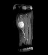

Findings: There is a large well-defined, oval-shape soft tissue lesion in the right upper medial thigh within the adductor longus muscle. The lesion is isointense to the muscles on T1 and hyperintense on T2 –weighted images. The lesion shows marked enhancement after the contrast administration. No significant cystic/necrotic changes, fat component, or calcifications are seen in it. No extra-muscular extension is seen. There is no invasion of myofascial planes, subcutaneous soft tissues, neurovascular bundle, or underlying femur. No skip lesion or significant lymphadenopathy is seen. Differential diagnosis includes neurofibroma, myxoma, or soft tissue sarcoma.
Case Discussion
the patient lost to follow up after initial sonographic and radiographic evaluation and 6 months later, presented with worsening symptoms. He underwent an MRI of the right thigh followed by a biopsy
histopathology: High grade undifferentiated pleomorphic sarcoma (previously known as malignant fibrous histiocytoma). Immunohistochemically, tumour cells react positively for Vimentin (V9) while negatively for Desmin (D33), SMA (1A4), Myo-D1 (5.8), S100 (polyclonal), CD34 (Qbend10), SOX10 (EP268), and PAN CK (AE1/AE3)


 Unable to process the form. Check for errors and try again.