
Presentation
Referred to the breast clinic with a lump in the left axilla.
Patient Data
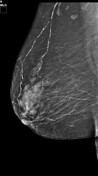

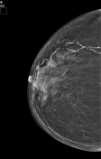

There are enlarged pathological lymph nodes seen in the left axilla.
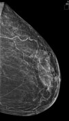
Incidental normal lymph nodes in the right axilla.
No mammographic evidence of a primary breast malignancy.
Incidental bilateral vascular calcification in both breasts.
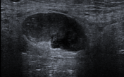
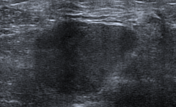

Ultrasound of the left axilla confirmed pathological left axillary nodes:
enlarged lymph node with a thickened cortex and near-complete effacement of the fatty hilum
pathological lymph node with a complete replacement of the fatty hilum
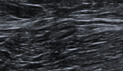
The normal contralateral axillary node with a reniform shape, fatty hilum, and cortical thickness <3 mm.
Case Discussion
In line with the principles of triple assessment following clinical examination and imaging, a core biopsy of one of the left axillary nodes was performed. The histology results confirmed metastatic signet ring cell adenocarcinoma with appearances and immune profile suggesting primary gastric origin.
The differential diagnosis of unilateral axillary adenopathy is broad and includes benign and malignant causes 1:
benign causes of unilateral axillary adenopathy include regional infection and silicone-induced granulomatous adenitis (in women with breast implants).
the most common malignant cause of unilateral axillary adenopathy is metastasis from ipsilateral primary breast cancer 1. Hence, imaging of the ipsilateral breast should always be performed 2. Other malignant causes include metastasis from non-breast primary malignancy (melanoma, thyroid cancer, lung, GIT, pancreas, ovary) and occasionally unilateral presentation of lymphoma.
Given this broad differential, a biopsy is necessary for a definite diagnosis.
This case demonstrates a rare presentation of metastatic gastric cancer with left axillary nodal metastasis.


 Unable to process the form. Check for errors and try again.