Presentation
Shortness of breath. Radiologic work up advised to rule out lung neoplasm.
Patient Data
Age: 70 years
Gender: Male
Download
Info

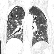
Decreased lung volumes are noted with coarse reticulation appears more pronounced peripherally and caudally.


Download
Info

- marked honeycombing bilaterally with traction bronchiectasis and lung volume loss
- reticular abnormalities bilaterally
- subpleural distribution with basal predominance
No feature very inconsistent with UIP pattern such as:
- ground-glass opacity outside areas of fibrosis
- mosaic perfusion/air trapping
- centrilobular nodules
- perilymphatic nodules
- subpleural sparing
- peribronchovascular predominance
Case Discussion
The features are representing a typical case of UIP pattern (definite) according to Diagnostic HRCT criteria for usual interstitial pneumonia (UIP) pattern - ATS/ERS/JRS/ALAT (2018).