
Presentation
Routine anomaly scan.
Patient Data
Age: 40 years
Gender: Female
From the case:
Uterine didelphys with pregnancy

Show annotations
Download
Info
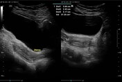
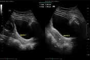
There are two uterine horns visualized, as well as two separate cervices. The left horn is non-gravid with a capacity of 37 cc and a cervical length of 1.59 cm.
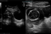
Single viable intrauterine fetus seen in the right horn in variable presentation with an AUA of 16 weeks 3 days and a regular fetal heart rate of 161bpm.
Fetal growth corresponds with a period of amenorrhea.
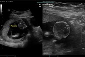
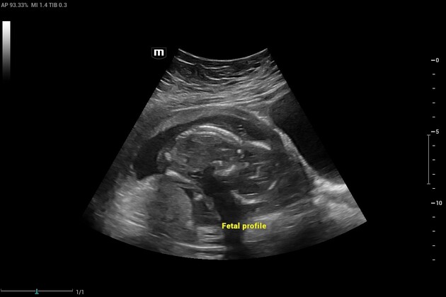
Normal fetal morphology. The placenta is fundal, well-attached, and not low-lying.
Amniotic fluid is adequate for gestation.
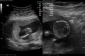
Cervix is closed and measures 5.3 cm in length.
Case Discussion
Uterine didelphys is as a result of Mullerian duct anomaly where there there are two separate uterine horns and two separate cervices.


 Unable to process the form. Check for errors and try again.