
Presentation
Difficulty in swallowing of one-year duration.
Patient Data
Age: 45 years
Gender: Female
From the case:
Vagal schwannoma


Download
Info

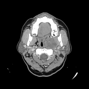
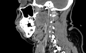
The CT images demonstrate a well-defined ovoid moderately enhancing soft tissue mass (4.2 x 3.7cm) within the left parapharyngeal space, compressing the oropharyngeal airway, and displacing the carotid vessels posterolaterally.
From the case:
Vagal schwannoma
Download
Info

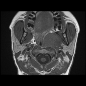
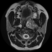
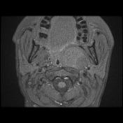
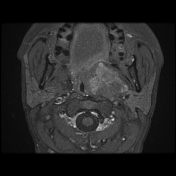
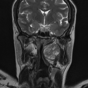
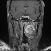
The previously described soft tissue mass appears well-encapsulated of intermediate signal intensity on T1WI, heterogeneous high signal on T2WI with moderate and heterogeneous enhancement following IV contrast.
Case Discussion
CT and MRI features are suggestive of a vagal schwannoma.


 Unable to process the form. Check for errors and try again.